クリストファー・ロータ博士
フローサイトメトリーの科学者
今月のFlow Mattersブログの記事へようこそ!Generating good flow data starts with picking the right mixture of fluorescent labels to use in your良好な流量データを生成するには、フローサイトメトリーアッセイ(一般的に「パネル」と呼ばれる) and the right type of flow cytometerおよび適切なタイプのフローサイトメーターで使用する蛍光ラベルの適切な混合物を選定することから始まる。When building flow assays, it helps tounderstand how flow cytometers readourmixtures of labels and producethe measurements that we spend so much time poring over during data analysisフローアッセイを構築する際、フローサイトメーターがラベルの混合物をどのように読み取り、データ解析中に長時間にわたって測定を行うかを理解するのに役立ちます。In this post, we’re going to do a deeper dive into the technical side of how biomarkerdetection in flow works and theこの記事では、フローにおけるバイオマーカー検出の仕組みの技術的側面と、実用的なアッセイ設計のための教訓についてさらに詳しく紹介します。
虹の向こう側――フローサイトの生体マーカー検出
As we talked about in our very first blog post, flow cytometersoperate on the principle that each label in apanel is attached to a fluorescent substance最初のブログ記事で述べたように、フローサイトメットは、パネル内の各ラベルが蛍光物質(fluorochrome一般的にはフルオロクロムとも呼ばれる)に結合しているという原理に基づいて動作する。These fluorochromes containone or more can これらの蛍光クロムには、サイトメーターのレーザーから特定の波長(色)の光を吸収し、別の波長の光を放出し、サイトメーターの検出器で捉えることができる、1つ以上の蛍光分子(蛍光体)from the cytometer’s lasers and emit light of another wavelengththat can be captured by the cytometer’s detectorsが含まれている。The precise patterns of absorption and emission for each fluorochromeform its excitation and emission spectra: a distinct set of behaviors that form a recognizable signature in themeasurements collected by our cytometerフッ素クロムの各クロムの吸収と放出の正確なパターンは、その励起スペクトルおよび発光スペクトルを形成する。すなわち、当社のサイトメーターが収集した測定において、識別可能な特徴となる異なる挙動である。The most important characteristics of this signature are the fluorochrome’speak excitation and peak emission wavelengths: the points where it most strongly absorbs and emits light, respectivelyこの特徴的な特徴として最も重要なのは、フッ素クロムのピーク励起波長とピーク発光波長であり、それぞれ光を最も強く吸収し、放出する点である。
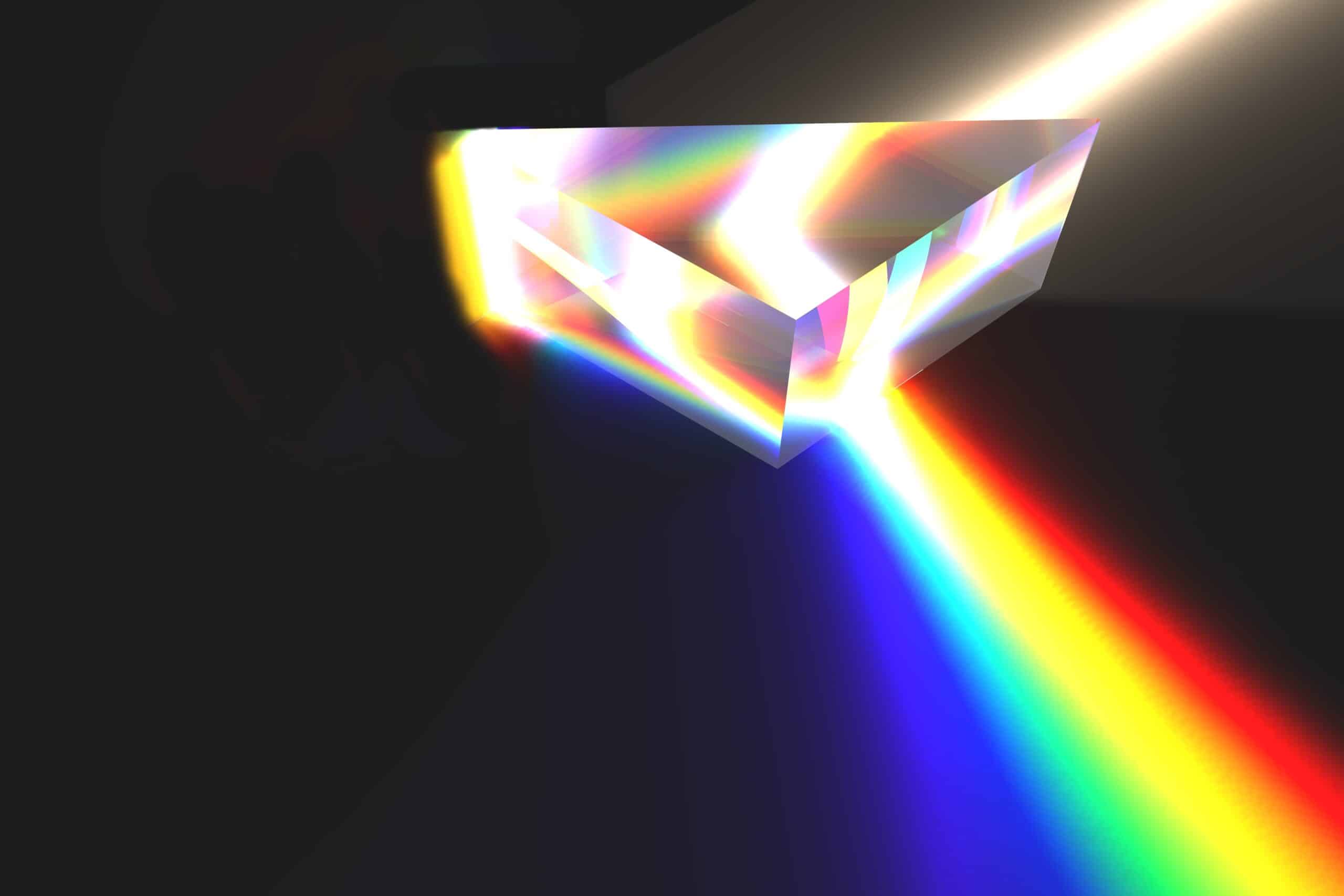
When picking reagents to use in a panel, it is important to use fluorochromes with as unique spectra as possibleパネルで使用する試薬を選ぶ際には、可能な限り独自のスペクトルを持つフッ素クロムを使用することが重要である。By pairing markers to labels with distinct fluorescent signatures, we can more accurately tracethe colors of light emitted by our sample back to theirマーカーを独自の蛍光署名を持つラベルと組み合わせることで、サンプルから放出される光の色をより正確にその由来(s (our labels and biomarkersラベルおよびバイオマーカーの関心)に遡って追跡できます。In a well-designed panel, a cytometer sees a cell like a human might see a rainbow: an array of carefully organized, unique colors that can be easily traced back to their originsよく設計されたパネルでは、サイトメーターが人間のように虹を見る細胞を見る。それは、その起源まで容易に遡ることができる、丹念に整理された独特な色の配列である。
With that image in mind, you might askその画像を念頭に置いて、虹の大きさ(何色)がサイトメーターを正確に「観測」できるかを問うべきだろう。答え:フローサイトメーターの構築方法によって異なります!
The first factor that influences a cytometer’s vision is how manyサイトメーターの視覚に影響を与える最初の要因は、レーザー(励起光の異なる色 of excitation light)itis equipped withがどれだけ備わっているかである。The more lasers an instrument has, the more options you have to excite your fluorochromes with, and the better you can separate them based on that behavior装置にレーザーが多く含まれているほど、フッ素クロムを興奮させるための選択肢が増え、その動作に基づいて分離する効果も高くなります。
As a simple example, let’s say we want to build a panel around two biomarkers: one marker for our cell type of interest (i.e CD3 for T cells) and one biomarker associated with the disease context we’re studying (i.e IL-17, an autoimmunity biomarker we discussed last time). We would expect both biomarkers to be found on the same cell at least some of the time, which means we need to pair them with highly distinct fluorochromes.
Two commonly used fluorochromes that could fit this bill are PE and APC. PE (phycoerythrin) can absorb light from blue or yellow-green lasers (reaching peak excitation at 565 nm) and emits a light red light (reaching peak emission at 578 nm). Allophycocyanin (APC) also absorbs yellow-green laser light but can uniquely absorb red laser light (peak excitation: 640 nm) and emits a darker red light (peak emission: 650 nm). If our instrument is equipped with both a yellow-green and a red laser, these two fluorochromes will be very straightforward to distinguish from one another. When we shine each laser on our cells in turn, we can be very confident that any signal we measure is coming from only our PE label or our APC label.
Using this same paradigm, we could expand our panel to include additional biomarkers using other distinctly excited fluorochromes, such as BV421 (best excited by violet laser light) and FITC (best excited by blue laser light). While there is some increased chance of misinterpretation for FITC, as PE also can be excited by blue light, we would expect the resolution of our two original biomarkers on PE and APC to be very similar between these 2 and 4 color panels. Success! We’re now collecting more information from our sample without any loss of accuracy – the dream scenario for any assay development scientist.
What happens if we want to add even more biomarkers, though? What do we do once we run out of lasers on our instrument?